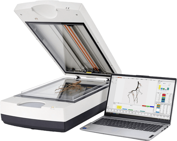
Plant Root Scanning System
- Product Number:LD-GX02S
- Product Difference:A3 Scanning-Based Root System Image Analysis
 +86 19353291814
 Online Consultation
Product Introduction
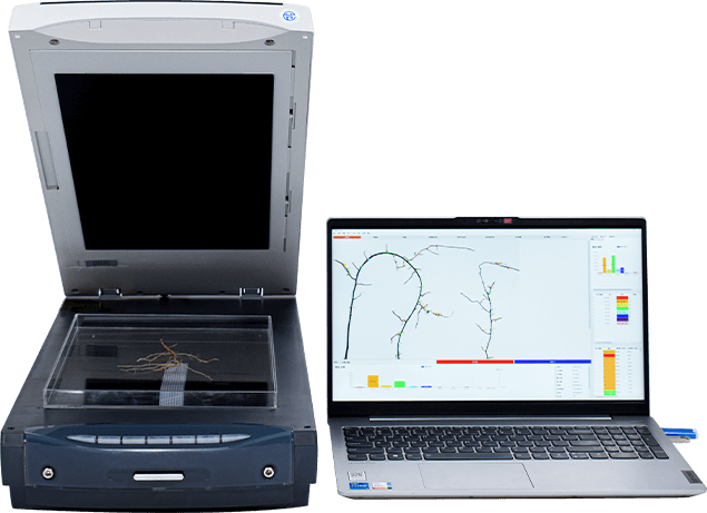
The plant root scanning system is used for professional root analysis after root washing and can also be used for root phenotypic analysis of plants grown in root trays. It can analyze root length, diameter, area, volume, root tip count, and more. It is powerful and easy to operate, and the software can analyze plant root morphology and the overall structural distribution of the root system. It is widely used in research on root morphology and structure.
The plant root scanning system uses a high-quality graphic scanner to capture high-resolution color or black-and-white images of plant roots. The scanner is equipped with a specialized dual-light source illumination system installed beneath the scanning panel and within the top cover, and the scanning panel features a dedicated dual-light source calibration area. Additionally, the system comes with specialized, highly transparent root placement trays of various sizes. During scanning, the light sources beneath the scanning panel and within the top cover simultaneously illuminate the root sample placed in the highly transparent root tray. This prevents the effects of shadows and unevenness that commonly occur during root scanning, effectively ensuring the quality of the acquired images.
This root analysis software can read images in standard TIFF and JPEG formats. For the acquired images, software that decrypts data using a dongle is used to analyze the high-quality root images obtained from scanning. Non-statistical methods are employed to measure and calculate basic morphological parameters—such as the length, diameter, area, volume, and root tip of overlapping root sections—thereby supporting researchers’ studies on different categories and levels of plant root systems.
Product Features
Product parameters
| Scanner | Equipped with an A3 dual-light-source color scanner featuring an optical resolution of 4800×9600. The scanning area for root reflection images is 304.8 mm × 429.26 mm, and for transmission scanning it is 304.8 mm × 403.86 mm, with a minimum pixel size of 0.005 mm × 0.0026 mm. |
| Measurements that can be analyzed | (1) Total root length; (2) Branching frequency; (3) Average root diameter; (4) Median root diameter; (5) Maximum diameter; (6) Total root area; (7) Total projected area; (8) Total root volume; (9) Number of root tips; (10) Number of forks; (11) Number of overlaps; (12) Parameters of root diameter grade distribution; (13) Customizable diameter segments with non-equidistant intervals; automatically measures the length, projected area, surface area, volume, and other parameters for each diameter segment, as well as their distribution parameters; (14) Capable of performing root system color analysis, outputting the diameter, length, projected area, surface area, and volume of roots of different colors; (15) Capable of performing root system topological analysis, automatically determining the number of root connections, relationship angles, etc., and can also automatically analyze the length, area, volume, and other parameters of any individual root segment separately; it can display the corresponding parameters for any diameter segment of a marked root system individually (with customizable, non-equidistant intervals); (16) Capable of performing root correction operations such as branch trimming, merging, and connecting; correction operations can be undone to quickly obtain 100% accurate results; (17) Capable of automatically measuring root fractal dimension using the box-dimension method. Can analyze the number of root nodules to objectively determine the contribution of rhizobia; (18) Supports fully automated root system analysis of large batches, with batch saving capabilities. Overall data can be summarized and saved in a single spreadsheet, or detailed data for each root system can be saved individually; (19) Extracts analytical characteristics for geometric angle and horizontal angle analysis; (20) All analysis images, distribution maps, and result data can be saved and exported to Excel spreadsheets; analysis annotation maps can also be exported; (21) The instrument is supported by a cloud platform, allowing analysis data to be saved to the cloud for viewing anytime, anywhere; (22) Built-in bilingual (Chinese and English) display with one-click switching for seamless integration; |
| Image Capture System Parameters | Scanning Element: 6-line interlaced microlens CCD; Maximum Format: A3; Interface Type: USB 3.1 Gen 1; Optical Resolution (dpi): 4800 × 9600 dpi; Maximum Resolution: 12800 × 12800 dpi; Minimum Pixel Size: ≥0.005 mm × 0.0026 mm; Scanning Light Source: White cold cathode fluorescent lamp (CCFL), 48-bit color depth; Scanning Area: 304.8 mm × 403.86 mm; |
| Optional | Brand-name computer (9th Gen or later Intel Core i5 CPU / 16 GB RAM / 21.5” color monitor / wireless network card, at least 4 USB ports including at least one USB 3.0 port, operating environment: Windows 10 Full Professional Edition or Ultimate Edition) |
 Prev
Prev Next
Next